Reliability of manually segmented anatomical landmarks in MRI scans to inform clinical gait analysis in patients with obesity
Aim and Research Question(s)
To provide added value and new research results in the field of gait analysis evaluation and therefore a validation tool for 3D freehand ultrasound based on segmentations of anatomical structures from MRI datasets, this master thesis is dedicated to answering the following research questions:
- What difference occurs in multiple manual segmentation of anatomical landmarks by the same operator?
- What is the intra-rater reliability error of manual segmentation of anatomical landmarks in MRI scans of obese children and adolescents?
Background
Gait and movement analyses are increasingly used as diagnostic and therapy-accompanying methods in connection with mobility restrictions [1]. Especially in the field of marker position determination in obese persons during the analyses, the demand for technical alternatives is increasing immensely [2]. A promising method for determining the position of retroreflective markers is 3D - freehand ultrasound, but a suitable validation instrument is required for this technique [3]. Manual segmentation of anatomical structures from MRI scans, provides the ability to do this.
Methods
To answer the research questions of this thesis, the left and right femoral head as well as the left and right spina iliaca anterior superior were manually segmented from 19 already collected MRI data sets of the pelvis of obese children and adolescents from a previous study of the University of Applied Sciences St. Pölten under the direction of FH-Prof. Priv.-Doz.Dr. Brian Horsak. The segmentations were repeated in three temporally separated measurement procedures. Through various mathematical calculations using Python, we obtained a visual representation of the femoral heads in the form of spheres scaled according to the collected data. Furthermore, the statistical results of the intraclass correlation coefficient (ICC), the t-test, the standard error of measurement (SEM), and the Bland Altman plot indicated the general reliability of the gathered data.
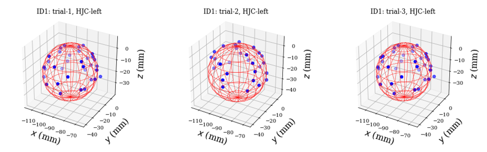 Figure 1: Visual Representation of the calculated Spheres
Results and Discussion
At a significance level of >0.05 and significance values between p= 0.074 and 0.742, no significant mean differences were detected between the femoral head segmentation trials. Similar results were obtained when calculating the mean differences for the medio-lateral (p = 0.087 - 0.999), anterio-posterior (p = 0.058 - 0.818), and inferior-superior (p = 0.110 - 0.696) positions of the ASIS segmentations. Using ICC (3, k), we obtained excellent values between 0.991 (SEM: ± 0.32mm) and 0.970 (SEM: ± 0.58 mm) for the segmentations of the femoral head radius as well as for the coordinate positions of the spina iliaca anterior superior with values between 0.990 - 0.999 (SEM: ± 0.87 - 2.40 degrees).
Conclusion
In summary, the results show that manual segmentation of anatomical structures from MRI Scans represents a suitable validation method for 3D freehand ultrasound determination of marker positions in the context of gait analysis.
References
[1]V. Stein und B. Greitemann, [2]V. Camomilla, A. Cereatti, A. G. Cutti, S. Fantozzi, R. Stagni, und G. Vannozzi, [3]Horsak, B., Schwab, C., Durstberger, S.