Development and usability evaluation of a 3D-printed heart model for biventricular pacemaker implantation practice
Aim and Research Question(s)
The aim of the master thesis was to develop a 3D cardiac model for biventricular pacemaker implantation practice. Medical professionals, including cardiologists, surgeons, and medical trainees, evaluate the model with regard to its usability. RQ 1: What is the workflow regarding the preparation of the 3D model starting from the CT dataset to a printed training model for a three-chamber pacemaker? RQ2: How do the subjects assess the usability of the 3D printed cardiac model for the placement of probes in a three-chamber pacemaker based on the results of the SUS and UEQ questionnaires?
Background
Heart failure, a leading cause of morbidity and mortality worldwide, decreases life expectancy, reduces quality of life, and increases health care costs. Although there are many different causes that can lead to heart failure, the primary cause is left ventricular systolic dysfunction. Insertion of cardiac resynchronization therapy (CRT) devices is significantly more complex than that of single- or dual-chamber systems and requires careful consideration and preparation [1]. The implanting physician must have a solid understanding of coronary vein anatomy, as the diversity of cardiac vein morphology can complicate successful insertion of a LV lead [2].
Methods
To facilitate training for the insertion of the left ventricular lead, a 3D printed model was developed using the Stereolithography (SLA) technique. Ten medical professionals specializing in cardiology, internal medicine, and surgery participated in the training. The effectiveness of the training was evaluated using the System Usability Scale (SUS) and the Utility Evaluation Questionnaire (UEQ).
Results and Discussion
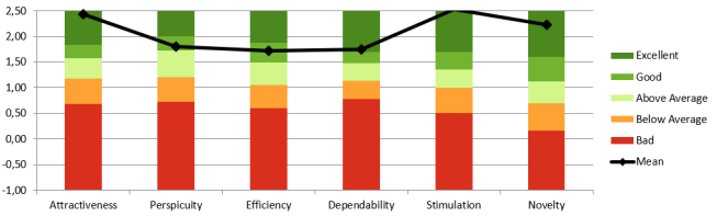
The evaluation of SUS resulted in an average score of 79.75, which is equivalent to a "C" grade according to Sauro [3]. The figure shows that the general user experience was rated positive in the UEQ. The values which were reached in the six subcategories where mainly rated “good” and
“excellent”. The values for the six attributes are the following: Attractiveness 2.43, Perspicuity 1.80, Efficiency 1.72, Dependability 1.75, Stimulation 2.52 and Novelty 2.22.
Conclusion
The creation of a 3D printed training model offers a comprehensive understanding of the heart's structure and its relevance to disease conditions. This knowledge benefits clinical practice by enhancing surgical procedures and transplantation techniques, as well as improving educational outcomes. However, it is important to acknowledge that processing CT data into a 3D model can be time-consuming and requires expertise in anatomy and technology. Additionally, the limitations of the model include the inability to accurately replicate tissue properties and simulate continuous fluid flow. Further investigation into contrast agent injection and radiopaque coating effects would be valuable.
References
[1] B. Osswald (2019), [2] G.-X. Yan et. al (2020), [3] J. Sauro (2012)